Epidermolysis bullosa in a lamb
Rachel Gibney – District Veterinary Officer
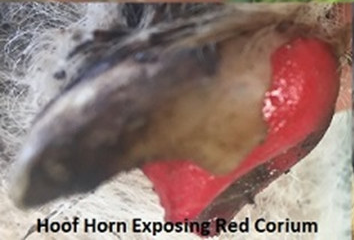
Towards the end of August 2018, a neonatal lamb was presented with one hoof detaching from the coronary band. The lamb originated from a small Wiltipoll flock on the outskirts of Ballarat. No other lambs in the flock were affected, including the twin of the lamb. Within 12 hours, a second foot was affected. The condition began with reddening of the coronary band and progressed to detachment of the outer hoof.
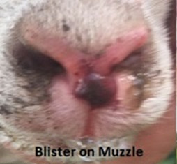
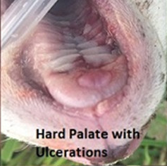
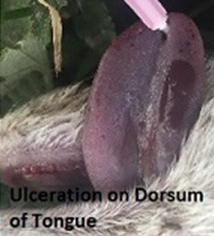
At 18 hours of age, a blood-filled blister developed on the muzzle. Multifocal ulcerations and raised bulla were observed on the hard palate, plus multifocal coalescing ulcerations on the dorsum of the tongue. The lamb was euthanised and samples sent to the AgriBio Veterinary Diagnostic laboratory for testing.


Histological findings indicated a dystrophic form of epidermolysis bullosa, also referred to as 'red foot disease'. This name arises from the exposure of the red corium of the foot once the hoof horn has detached. Histology showed multifocal separation of the epidermis from the dermis, with areas of infiltration by macrophages, capillary hyperplasia and superficial proteinaceous exudate.

Separation occurs just below the basement membrane, where anchoring fibrils hold the basement membrane to underlying collagen. A defect in the protein leads to a deficient number of anchoring fibrils being present, and loss of adhesion between the dermis and epidermis.
Epidermolysis bullosa has been reported in numerous breeds of sheep including Corriedale and Suffolk sheep in Victoria. The cause is idiopathic, although a hereditary component is suspected. Skin may slough from the limbs and pinna, as well as the epidermal layers of the cornea.
No treatment options are available, and if not euthanised the lambs will succumb to secondary infections, or complete sloughing of the hooves, as was occurring in this lamb.