How to remove a brain in sheep, goats and cattle
WARNING: Some photos on this page could offend some readers. This graphic representation is necessary to allow for the procedure described below to be accurately performed.
Laboratory examination of the brain for transmissible spongiform encephalopathies (TSEs) requires its removal from the skull (cranium), whole and intact, and without damage to the brainstem.
Longitudinal craniotomy involves splitting the skull (but not the brain) ventrally and dorsally along its longitudinal axis with an axe.
The axe can be hit with a small sledge hammer for greater control and safety. Eye protection should be worn.
The two halves can be levered open from the front end to expose the intact brain. This method requires practice. It has the advantage of exposing the pituitary gland. The technique is similar for sheep and cattle.
The procedure can be performed quickly, simply and safely under field conditions with minimal equipment.
Method of removal
Figures 1 to 9 illustrate the removal of a sheep brain. The equipment required includes:
- an axe
- small sledge hammer
- boning knife
- disposable rubber gloves
- safety glasses
- hedge shears or similar
- personal protective equipment appropriate to the clinical case.
Removing the head
Bend the head back to extend the neck and commence severing the ventral neck at the level of the atlantooccipital joint. When the cervical spinal cord is exposed within this joint, sever the cord at the most caudally accessible site to leave 2 to 3cm of spinal cord attached to the brain stem.
Preparing the head
Skin the ventral head and remove the tongue and soft tissues of the throat so as to expose the hard palate and ventral cranium (Figure 1).
Hedge shears may be needed at this point to cut the hyoid apparatus.
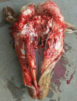
Using an axe, split the front of the bottom jaw, crack the cranium along the midline and split the hard palate and nose to full depth.
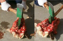
Turn the head over and cut the skin on the top of the head along the midline. Extend the cut deeply into the soft cartilage of the nose in the midline to split the nose (Figure 3).
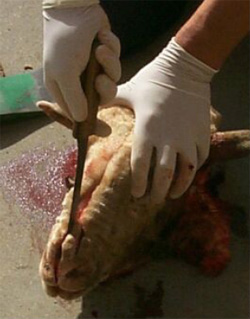
To help stabilise the head the 2 sides of the mandible can be moved laterally to provide a wide base.
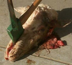
Splitting the skull
With an axe, crack the dorsal skull along the length of the midline where the skin cut has been made. The axe can be hit with a small sledgehammer for better control (Figure 5).
If the animal was euthanased by the use of captive bolt the penetration hole can provide a good starting point for the axe.
The aim is to crack the bone (cranium) surrounding the brain without damaging the brain but fully cut through the depth of the nose and jaw.

Levering the head open
With a knife, cut any soft tissue attachments that might prevent the head being levered apart (using the split nose).
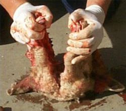
Grab the halves of the split nose and slowly prise and pull them apart (Figures 6, 7). More bone cracking will be required if the head won't come apart easily. If the cranium has been cracked sufficiently, the whole head can be levered open and the brain and pituitary gland exposed.
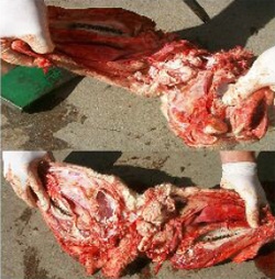
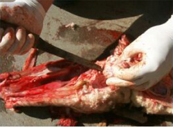
A boning knife (or scissors) is used to cut the nerve roots and dura mater as the brain is exposed and removed (Figure 8). The brain can be removed whole and intact.
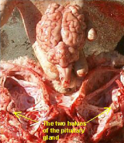
The hemisected pituitary gland is exposed at the base of the brain (Figure 9). It is easily removed if required.
Remove 2 to 3cm of the spinal cord and place in a sterile container. Ensure the obex is left intact.
For sheep and goats TSE surveillance the top third of the cerebellum must also be placed in the sterile container with the spinal cord. If these samples cannot be sent to the laboratory on the day of removal they should be frozen.
Place the whole brain in formalin, brain stem up to prevent pressure on the tissues of interest.
Common mistakes
Some common mistakes are:
- cracking (cutting) too deeply into the bone around the brain, particularly the bone protecting the ventral brain, can damage the brain stem
- insufficient 'cracking' of the bones surrounding the brain, particularly the ventral cranium, can make it difficult to lever the cranium apart. One side of the nose breaks.
- levering the head apart too quickly can tear the brain.